
.webp)
A Freak Accident, a Serious Injury
When Ralphie, a 17-week-old golden-black Cockerspaniel cross, jumped from a bicycle basket and became entangled in the front wheel, the result was a severe combined fracture and dislocation injury to his left hind leg.
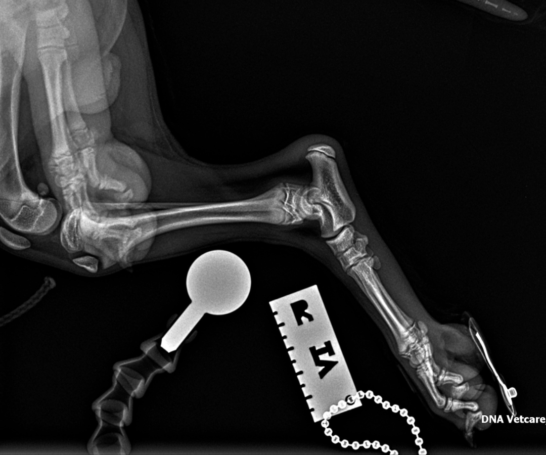
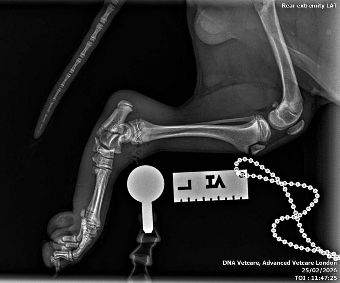
Thanks to the quick thinking of Ralphie’s owner, Amy, who immediately took him to their primary veterinary surgery The Vet on Richmond Hill, he was referred to Advanced Vetcare London the following day. Ralphie arrived unable to bear any weight on the injured leg, with significant swelling around the tarsal (ankle) joint and visible skin trauma. Behind those clinical signs lay a picture that specialist imaging would confirm as far more complicated than just a straightforward fracture.
The Diagnosis: Multiple Fractures and Dislocations
X-rays and cone beam CT of Ralphie's left hindlimb - a technique that uses a cone-shaped beam to produce detailed 3D images, particularly useful in complex orthopaedic cases like this - revealed a combination of injuries that, together, posed a serious surgical challenge:
- A comminuted fracture of the base of the left calcaneus (heel bone), meaning that the bone had broken into multiple fragments
- A small avulsion fracture in the calcaneoquartal region, whereby a small piece of bone had been pulled away from the joint when it was suddenly pulled by the long plantar ligament
- Dislocation of the talo-calcaneal joint - the lower ankle joint that connects the top ankle bone to the heel bone
- Plantar and lateral dislocation of the talocentral joint - this meant that the ligaments holding the bones in the hind leg together had completely torn, and the main hinge joint had broken, causing the paw to collapse toward the ground (plantar) and shift outward toward the side (lateral).
The severity of the injury, particularly the ligamentous disruption alongside the fractures, meant that non-surgical, non-invasive management was not a viable option. Surgical stabilisation was the only path to restoring Ralphie's limb function.
The Surgery: Rebuilding the Ankle
At just 4 months old, this added another layer of complication to the procedure to stabilise Ralphie's leg. A puppy’s growth plates don’t fully harden until around 18 months of age. This means that operating on a puppy's developing skeleton, with multiple unstable fragments and disrupted soft tissue, demands both technical precision and the ability to adapt when anatomy doesn't cooperate. Ralphie's procedure required exactly that.
Stabilising the Fractures
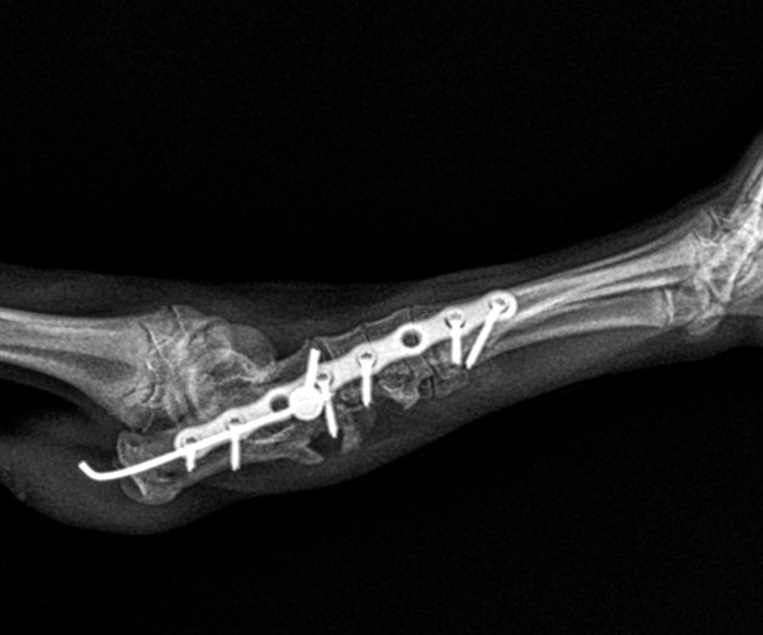
The first priority was bringing the broken heel bone back into alignment. A fine pin (1.1 mm intramedullary pin) was passed retrogradely through the calcaneal fragments, essentially acting as an internal scaffolding rod, holding the pieces together while the more complex reconstruction took place around them.
With the heel bone provisionally stable, the team turned their attention to Ralphie’s dislocated joints. Because several of the small tarsal joints had been thrown completely out of position, they needed to be temporarily bridged and held still to allow the disrupted ligaments time to heal. A slim, precision locking plate (a 1.5 mm 8-hole RL Leilox plate) was secured along the outer side of the ankle region. This specialised implant bridged across the small bones of the hock joint, running from the metatarsal (foot) bones, through the central tarsal and fourth tarsal bones in the ankle, and anchoring into the calcaneus (heel bone). This provided stability across the affected area.
Locking screws anchored at multiple points meant that the load was distributed across the repair rather than concentrating stress at any single fixation point, which is particularly important in a puppy's developing bone. An additional cortical screw fitted with a small washer was inserted through the lower fragment of the calcaneus (heel bone) and into the talus (the bone sitting directly above the heel) to further strengthen and stabilise the overall repair.
Repairing the Ligaments
One of the more technically demanding aspects of Ralphie's case involved a small, flake-like bone fragment at the centroquartal joint - the junction between the central and fourth tarsal bones. Here, the long plantar ligament had partially torn away. Because the fragment was too small and delicate to hold a conventional screw or pin, stabilising it with standard hardware was not an option.
Instead, the surgical team reattached the ligament by passing sutures through small tunnels carefully drilled into the main body of the heel bone. This tunnel technique restores the critical load-bearing and stabilising function of the plantar ligament while avoiding the risk of fragmenting tissue that simply cannot support metallic implants.
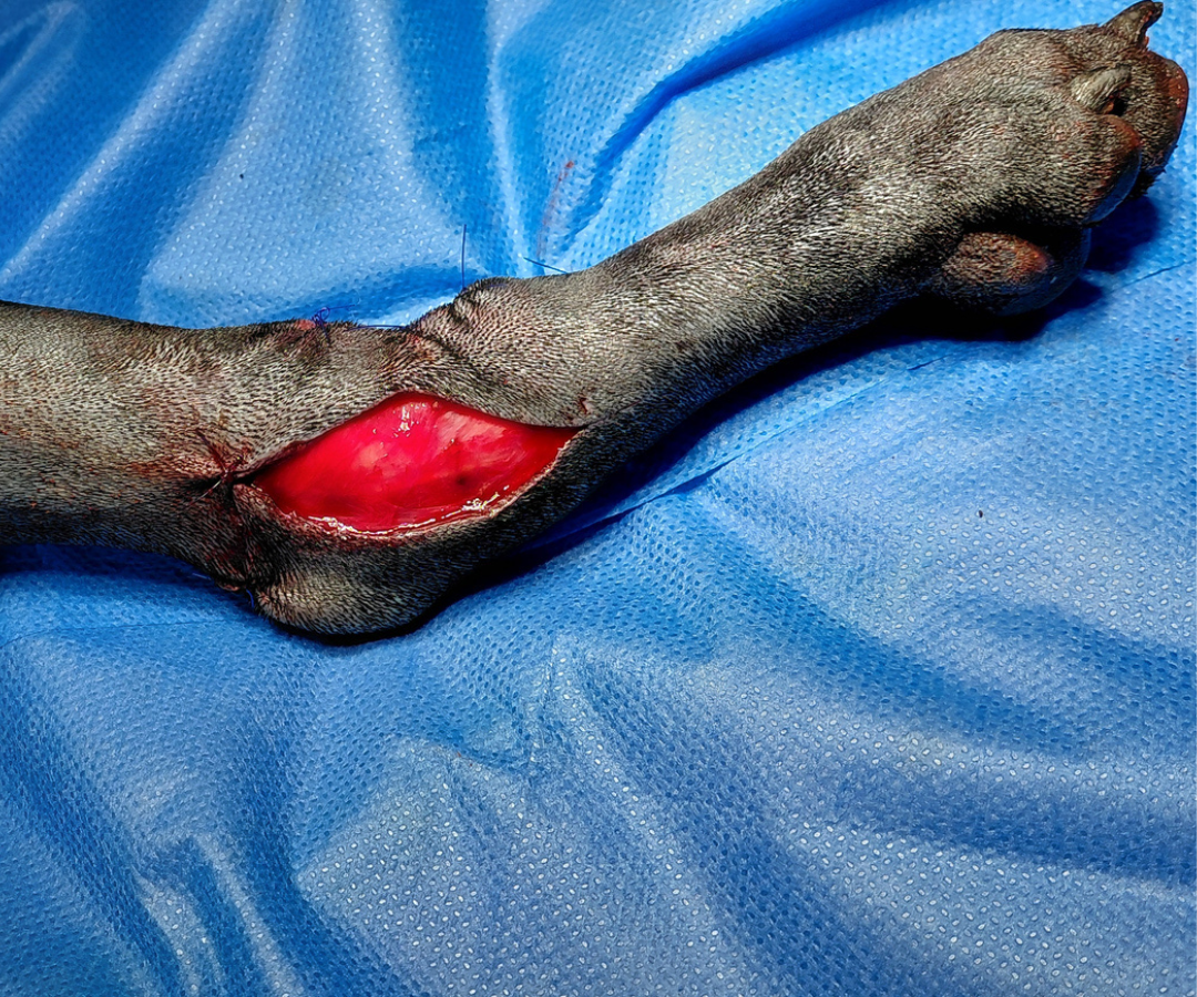
Managing the Soft Tissue Wound
An additional challenge was Ralphie’s de-gloving injury where the skin around his main ankle joint had been stripped away from the underlying tissue. The wound was surgically cleaned and cleared of any non-viable tissue (debrided) and a carefully placed incision along the inner aspect of the leg was made to allow the surrounding skin to be drawn together without undue tension.
The open area was then dressed with a combination of medical-grade manuka honey, which supports natural wound healing and helps control infection, and Allevyn foam for gentle cushioning and moisture management. Dressing changes were planned every one to two days, guided by the level of wound discharge.
Post-operative X-rays confirmed that the fractured bones had been successfully realigned and that all implants were correctly positioned.

Recovery at Home
Ralphie was discharged the same evening, going home with his owner with clear instructions to support his recovery:
- Strict cage rest, with only 5 - 10 minute lead walks, three to five times daily
- Elizabethan collar worn at all times to protect the wound
- Regular bandage changes to manage the open wound until fully healed
- Skin sutures to be removed at 14 days
- Medications to manage pain (particularly nerve pain) and inflammation
Five Weeks Later: Full Healing, Implants Removed
When Ralphie returned to Advanced Vetcare London five weeks later, the transformation was remarkable.
X-rays confirmed that his fractures had completely healed. On examination, Ralphie was bright, alert and showing only mild lameness in the affected leg - an outstanding result given the severity of the original injury.
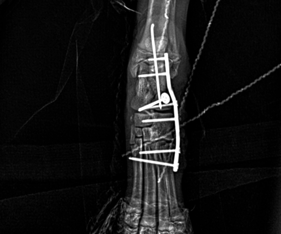
With the fractures healed and ligament structures confirmed to be stable, the decision was made to proceed with the removal of the plate, screws and pins
Postoperative radiographs confirmed complete implant removal and maintained fracture healing. Ralphie went home that evening with a light protective bandage and a short course of anti-inflammatories. His next visit is in a week for bandage review, with suture removal at 14 days.

What Makes This Case Stand Out
Ralphie's case is a reminder that severe orthopaedic injuries in puppies, while daunting, can be managed to a full recovery when diagnosis is thorough, surgical planning is meticulous, and the team is prepared to adapt their technique to what the anatomy demands.
Several elements of this case are worth highlighting:
Cone beam CT for precise surgical planning.
Standard X-rays were enough to identify the fracture, but CT imaging allowed the team to build a far more complete picture before entering the operating theatre. That level of detail directly determined which strategy was chosen and where each implant was placed.
Ligament repair through bone tunnels.
When a bone fragment is too small and fragile to hold a screw or pin, an alternative approach is needed. By threading sutures through small tunnels drilled into the main body of the heel bone, the surgical team were able to reattach the torn ligament and restore its stabilising function - an outcome that would not have been achievable with conventional hardware alone.
Temporary joint bridging as a means to an end, not a permanent solution.
The locking plate was always intended as a temporary measure. By holding the affected joints rigidly in place, it gave the ligaments and fractured bone the protected environment they needed to heal. The result was a repair stable enough for full implant removal and five weeks after surgery, Ralphie's joint was functioning without any hardware at all.
Proactive soft tissue management.
Ralphie’s skin wound was addressed from the outset with appropriate wound dressings and a carefully placed release incision to reduce skin tension. This allowed the wound to heal cleanly over the plate within approximately two weeks, which was a critical factor in keeping infection away from the implants during the recovery period.
To refer a case or find out more, visit advancedvetcare.co.uk or call 020 3143 4444.