
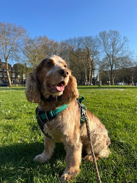
Mash, a nine-year-old male Cocker Spaniel, was referred to the Neurology and Neurosurgery Service at Advanced Vetcare London with sudden, severe neck pain that had not responded to a week of conservative treatment. Our investigation revealed an acute intervertebral disc extrusion between the fifth and sixth cervical vertebrae in the lower neck, so Mash went on to have same-day ventral slot surgery.
The Diagnosis
Mash had been suffering with cervical pain and stiffness for over a week when he was referred. He had already started conservative management with metacam, with no improvement, and the bloodwork had come back unremarkable.
Mash was quiet and reluctant to move, held his head low, would not turn it and was visibly trembling with pain. He was ambulatory but his gait was stiff and stilted. Reassuringly, paw placement and segmental spinal reflexes were normal in all four limbs, pointing towards a localised problem in the neck rather than more widespread spinal cord dysfunction.
An MRI scan of the cervical spine confirmed the cause: a moderate volume of degenerated and mineralised disc material within the vertebral canal at C5-C6, causing moderate to marked compression of the spinal cord. The diagnosis was a C5-C6 intervertebral disc extrusion, a condition which develops when disc material degenerates and calcifies before rupturing suddenly into the canal.
The Treatment
Mash underwent a single general anaesthetic covering both the MRI scan and surgery, performed back to back. Prof. Dr. Marios Charalambous and his team carried out a C5-C6 ventral slot: a window cut through the floor of the vertebral canal from beneath the neck, giving direct access to the compressive material without needing to manipulate the spinal cord itself. The degenerated disc material was removed from the canal. The spinal cord was then visualised and appeared normal, with no signs of ongoing compression or damage.
“Cervical disc extrusions can look frightening when a dog first comes in,” explained the surgeon, Prof. Charalambous, “but cases like Mash’s are a good reminder that a nine-year-old dog with a well-localised neurological condition still has an excellent chance of getting back to normal.”
Recovery
Mash recovered well from the anaesthetic and remained hospitalised for close monitoring over the following few days. He had occasional episodes of pain and vocalisation but was otherwise comfortable, moving his head and neck freely. The team felt some of this residual reaction was likely down to a combination of a low pain threshold and a behavioural component rather than ongoing surgical pain, something discussed openly with Mash's owner.
By the time he was discharged, his neurological examination was normal and he was showing no signs of neck pain or stiffness.
Mash's home care plan reflected a genuinely multimodal approach to pain relief: Pardale-V tabs, 100mg Trazodone and 750mg Methocarbamol for one week; 400mg Gabapentin and Metacam solution for three weeks and, finally, 100mg Amantadine, for four weeks. These helped manage the pain from several angles.
Strict rest was essential too. Mash's owner was asked to keep him confined to a playpen or small room for four weeks, with no jumping, stairs, furniture or play. Exercise was to be on a harness and lead and only for toilet breaks.
Raised food and water bowls meant he did not need to lower or bend his neck to eat and drink while it healed.
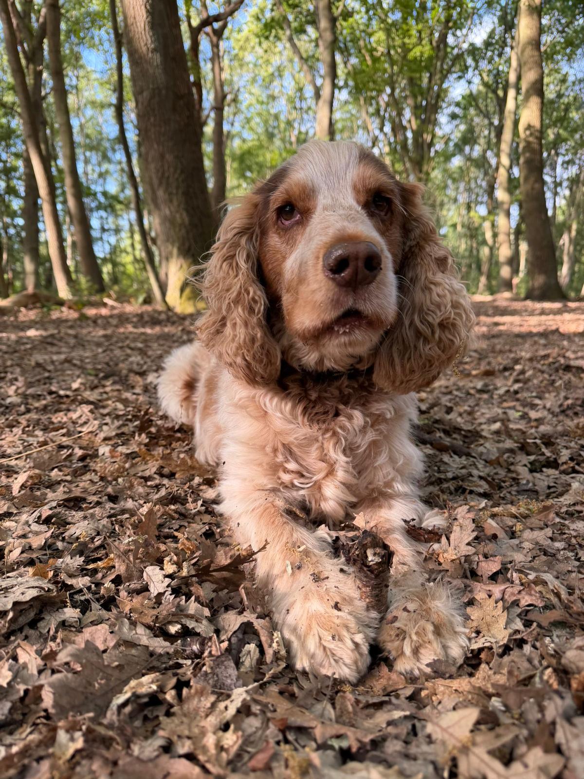
Four weeks on, Mash returned for his recheck and the improvement was clear. His owner reported he was back to himself. On examination his mentation, gait and stance were all normal, his postural reactions were intact and there was no pain on palpation or movement of the neck. His surgical wound had healed well.
He was cleared to progressively increase his exercise over the following month, avoiding running, jumping, stairs and slippery floors in the meantime.
What Makes This Case Stand Out
Cocker Spaniels get cervical disc disease too
Cervical intervertebral disc extrusion is often thought of as a Dachshund problem, but Mash's case, and the wider literature, show Cocker Spaniels present with it as well, typically later in life than more classically chondrodystrophic breeds. Age alone should not rule cervical IVDE in or out (Abouzeid, et al, 2025).
Recurrence is a real but manageable risk
A recent study following dogs after cervical ventral slot surgery found recurrence in around a quarter of cases over a median follow-up of almost four years, most commonly at an adjacent disc space, and found prophylactic fenestration of neighbouring discs reduced recurrence substantially in their cohort (Berthomé et al, 2025).
This is not a reason to avoid surgery given the alternative of ongoing compression, but it is worth factoring into long-term conversations with owners about monitoring for new signs.
Pain expression is not always a simple reflection of tissue healing
Mash's occasional vocalisation and stiffness after surgery, despite a technically successful procedure and a normal-appearing spinal cord, is a reminder that pain after major spinal surgery can have a behavioural as well as a nociceptive component. A multimodal plan, addressing both, gave Mash the best chance of a comfortable recovery.
Full recovery is achievable and fast
At his four-week recheck Mash was pain-free, moving normally and needing no further medication. For an older, medium-sized dog with marked spinal cord compression on presentation, that is an excellent outcome and a strong argument for timely surgical referral once conservative treatment has failed.
To refer a case or find out more about our neurology and neurosurgery services, you can contact us here, or call 020 3143 4444.