

How It Started
Loki is an outgoing, sociable cat who has spent his ten years making the most of South London life. So when his owners noticed he had gone off his food and spotted an unusual lump protruding from his belly, something clearly wasn't right.
He was seen by the team at Advanced Vetcare London's Streatham Hill sister practice, where a CT scan and tissue sampling were performed. The results pointed to a tumour of the left kidney, with cytology suggesting renal carcinoma.
The encouraging news was that the right kidney appeared to be functioning well. That opened up a clear surgical option - remove the affected kidney along with the tumour, and give Loki the best possible chance of a healthy life on one.

The Surgical Challenge
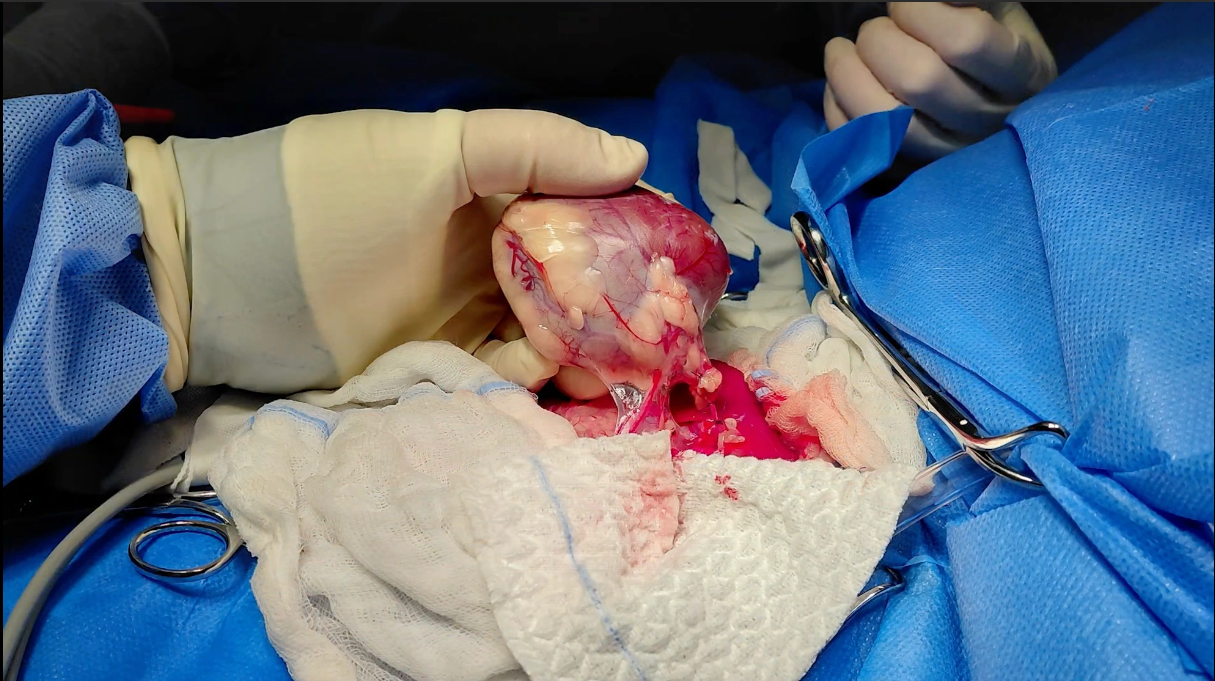
Nephrectomy (the surgical removal of a kidney) is never a straightforward procedure, but in Loki's case the size of the tumour added a significant layer of complexity.
Pre-operative planning is everything in a case like this. The CT scan gave the surgical team a detailed picture of the tumour's size and position before Loki went into theatre - crucial information when operating close to some of the body's most important blood vessels. The tumour appeared contained within the kidney capsule, which was an encouraging sign that it hadn't spread beyond the organ itself, meaning that if the kidney could be removed cleanly, there was a real chance of achieving clear margins.

"There are some critical steps in performing a nephrectomy," explains Mr. Jakub Köcher-Vodnárek, soft tissue surgery specialist at Advanced Vetcare London. "You have to make sure you ligate all the veins. Cats can have more than one draining into the kidney and there's very little margin for error when you're working close to major vessels like the aorta and caudal vena cava."
Because of the tumour's size, an open surgical approach was used to give the team the space and visibility needed to work safely. The kidney was carefully mobilised before the renal artery, renal veins and ureter were ligated, and the kidney and ureter removed together as a single specimen. The ureter was tied off close to the bladder as part of the same procedure.
In procedures this close to major blood vessels, specialist equipment makes a meaningful difference. Advanced Vetcare London's access to a vascular stapler - a tool that allows precise, secure closure of large vessels - was available throughout, offering an additional layer of safety during the most technically demanding stages of the surgery.
After Surgery
Loki came through the procedure well. In an encouraging early sign, he ate something shortly after waking from anaesthesia which is always a positive indicator in cats following major abdominal surgery.
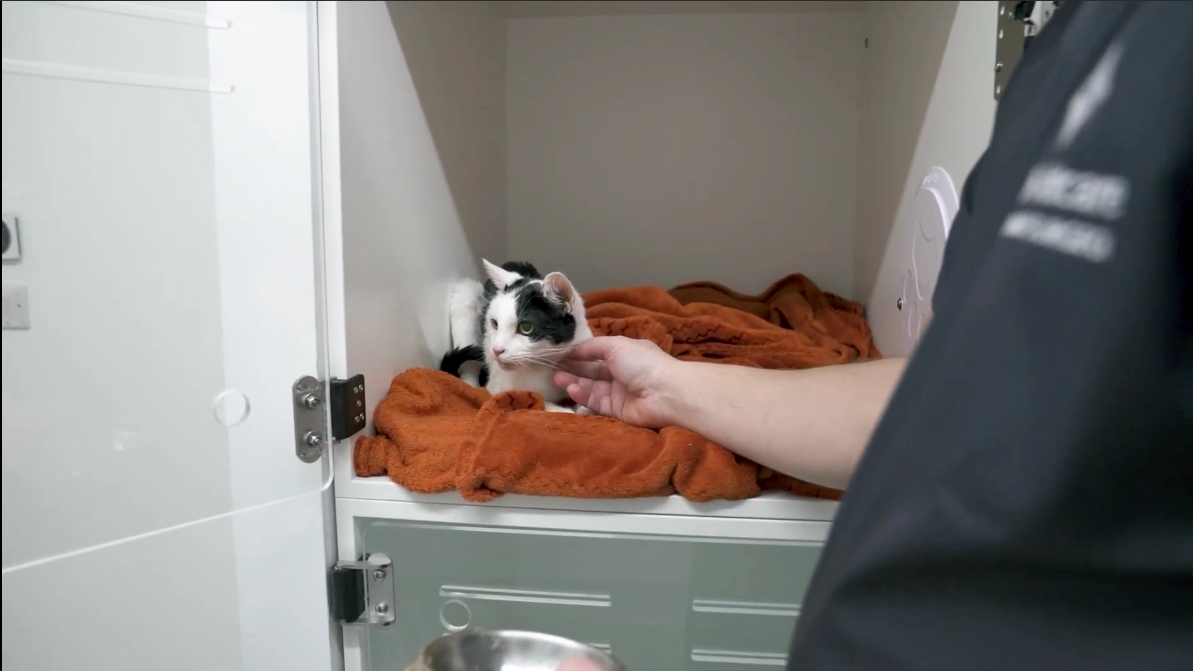
He remained hospitalised overnight for monitoring and supportive care, receiving intravenous fluids to support his remaining kidney as it adjusted to doing the work of two. Blood tests the following day allowed the team to assess how his kidney values were responding, a crucial checkpoint after any nephrectomy. Over the days that followed, Loki's appetite steadily improved, and he remained bright, alert and comfortable in his kennel.
Going Home
Once Loki was stable, he was discharged with a carefully structured plan to carry him through recovery at home. Subcutaneous fluids administered twice weekly helped support kidney function in the early weeks, alongside a renal diet designed to reduce the workload on his remaining kidney. Pain relief and anti-nausea medication kept him comfortable and eating well, and strict cage rest for a month gave his surgical site the time it needed to heal fully.
Follow-up blood tests were scheduled for approximately a week after discharge, the most important early checkpoint to confirm his remaining kidney was coping well.
What This Case Teaches Us
Loki's case is a good reminder that a cancer diagnosis, even in an older cat, doesn't always mean the end of the road. When a tumour is confined to a single organ and the other is functioning well, surgical removal can be genuinely life-changing. And, in some cases, curative.
It also illustrates why thorough pre-operative diagnostics make such a difference. The CT scan and cytology performed before referral meant the surgical team went into theatre with a clear picture of what they were dealing with, allowing for careful planning rather than intraoperative surprises.