

Advanced Vetcare London successfully treated a complete cranial cruciate ligament rupture in a 9-year-old Jack Russell Terrier cross using arthroscopy and TPLO surgery.
A Sudden Lameness After a Walk in the Park
Frank is a 9-year-old neutered male Jack Russell Terrier cross. He is the kind of dog who has no idea he's nearly a decade old! So when his owner noticed he was struggling to bear weight on his left hind leg after a run in the park, it was a worrying change.
Frank had already been seen at his regular practice and prescribed pain relief, but there was no improvement over five days. His owners decided to seek a second opinion, and he was referred to Advanced Vetcare London.
On examination, the picture was clear. Frank was completely non-weight bearing on the left hind limb. His stifle (knee joint) was markedly unstable, with both a positive tibial compression test and a positive cranial drawer sign detectable on conscious examination. These are two of the most reliable clinical indicators of a cranial cruciate ligament (CCL) rupture, the canine equivalent of an ACL tear in people.
The Diagnosis: Complete CCL Rupture
The cranial cruciate ligament is one of the primary stabilising structures of the dog's stifle joint. When it ruptures completely, the tibia (shin bone) is able to slide forward relative to the femur (thigh bone) which is exactly what the cranial drawer and tibial compression tests detect. Left untreated, a complete CCL rupture leads to progressive joint instability, pain, and arthritic change.
In Frank's case, surgery was needed.
Given Frank's strong clinical signs, radiographs were deferred to the day of surgery rather than taken at the initial consultation, avoiding an unnecessary extra visit and keeping things as straightforward as possible for Frank and his owners.

What the Radiographs Showed
Pre-operative X-rays of the left stifle confirmed increased joint effusion (fluid build-up, consistent with inflammation from the rupture) and mild osteophyte formation around the patella and fabellae. These are early arthritic changes that are common in dogs with CCL disease. The tibial plateau angle (TPA) measured 30 degrees, which is relevant to surgical planning for TPLO.
Interestingly, there were also mild changes visible in the right stifle and both hips. These weren't causing Frank problems at the time, but they're worth keeping in mind for ongoing monitoring.
The Surgery: Arthroscopy and TPLO
Frank's procedure combined two techniques: arthroscopy and TPLO.
Arthroscopy
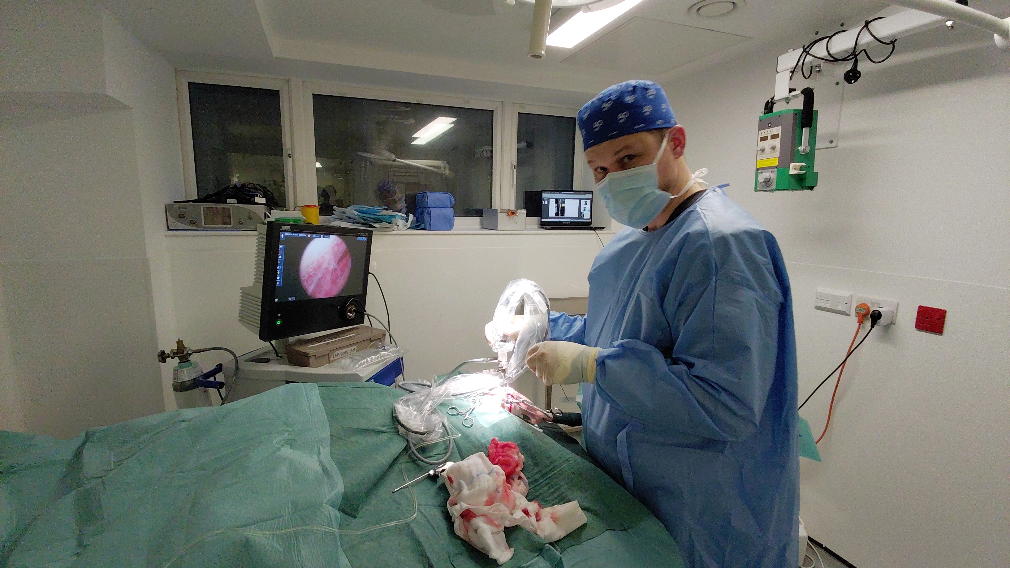
Before moving to the corrective bone surgery, the joint was assessed arthroscopically - a keyhole technique using a 1.9 mm 30° arthroscope with an extra-articular distractor to improve visualisation. This confirmed the complete CCL rupture directly and, importantly, showed that both the medial and lateral menisci were intact. The menisci are cartilage pads that act as shock absorbers within the stifle, and meniscal damage is a common complicating factor in CCL cases. Finding them intact was a good result for Frank.
TPLO
TPLO - Tibial Plateau Levelling Osteotomy - is currently the gold standard surgical treatment for CCL rupture in dogs. Rather than attempting to replace the ligament directly, the procedure works by changing the geometry of the joint itself. A curved cut is made in the top of the tibia and the tibial plateau is rotated to reduce its angle. This eliminates the forward sliding force (tibial thrust) that the CCL normally resists, effectively making the joint stable without needing the ligament at all.
In Frank's case, a 15 mm crescentic saw blade was used to make the osteotomy. The tibial plateau was rotated 6.7 mm and stabilised with a 2.4 mm VI TPLO plate. Intraoperative tibial compression testing confirmed that cranial translation had been eliminated. In other words, the instability that had been causing Frank's lameness was gone.
Post-operative radiographs confirmed appropriate osteotomy position and correct implant placement. Frank recovered from anaesthesia without any issues and was discharged the same evening.
Mr. Jakub Köcher-Vodnárek, Diplomate ECVS, Advanced Vetcare London
“There's sometimes a hesitation around putting older dogs through orthopaedic surgery and I understand that. But Frank is a great example of why age alone shouldn't be the deciding factor. A complete CCL rupture doesn't get better on its own, as the joint keeps deteriorating. With the right surgical technique and a sensible recovery plan, dogs like Frank do incredibly well. Seeing him walk out at eleven weeks with no lameness and no medication was amazing.”
Recovery at Home
Frank went home with a structured recovery plan:
- Pardale V: 1 tablet twice daily for 2 weeks
- Gabapentin 100 mg: one capsule three times daily for 5 days
- Elizabethan collar until suture removal at 14 days
- Strict cage and pen rest with lead-controlled exercise only for 8 weeks, starting at 5-10 minutes three to five times daily and increasing by 5 minutes per week
- Physiotherapy with an ACPAT chartered physiotherapist recommended
- Orthopaedic recheck at 8 weeks post-operatively
11 Weeks Later: Back to Full Function
When Frank came back for his recheck at 11 weeks post-operatively, the progress was excellent.
His owner reported that Frank had returned to normal, with no lameness and no pain relief needed. Also, he was comfortably managing walks of up to an hour and a half, which, for a Jack Russell cross, is exactly as it should be.
On examination, there was no swelling or discomfort in the left stifle, and gait assessment showed no lameness at all.
Given Frank's complete clinical recovery, the question of follow-up radiographs was discussed openly with his owner. Current evidence - including Alexander et al. (2021) - suggests that in cases of full clinical recovery, routine post-operative radiographs may not add meaningful clinical value. The owner elected not to proceed with imaging, and Frank was discharged.
What Makes Frank's Case Stand Out
Frank's recovery is a good example of what TPLO can achieve, even in a dog approaching his tenth year. A few things are worth highlighting.
Age is not a contraindication. At 9 years old, Frank is not a young dog. But CCL rupture in older, smaller breeds responds extremely well to TPLO, and his outcome reflects that. The key is thorough assessment, including the mild changes seen in his other joints, and setting realistic expectations with the owner from the outset.
Arthroscopy adds diagnostic confidence. Performing arthroscopy before the TPLO allowed direct confirmation of the diagnosis and critically, meniscal assessment. Knowing the menisci were intact before making the osteotomy meant the surgical plan could proceed without additional intervention.
Evidence-based decision-making around aftercare. Not every case needs the same post-operative workup. Frank's owner was given a clear, honest conversation about the current evidence on routine post-op radiographs in fully recovered patients, and together they made a pragmatic decision. That's how it should work.
Full function, no ongoing medication. At 11 weeks, Frank needed nothing. No anti-inflammatories, no pain relief, no restrictions. Just a dog getting back to his walks.
To refer a case or find out more about our orthopaedic services, visit advancedvetcare.co.uk or call 020 3143 4444.