
Dolly, a 14-year-old domestic shorthair cat, arrived at Advanced Vetcare London in a critical condition. A blocked ureter, a severely infected kidney, and the imminent risk of renal rupture meant surgery couldn't wait. What followed was a complex emergency ureteronephrectomy that gave this elderly cat her life back.
How It Started
Dolly's owners first noticed something was wrong when she stopped eating properly and began urinating in unusual places around the house. She had been gradually losing weight, not dramatically, but enough to be noticeable. By the time she was seen at her local vets, she was very weak.
Blood tests showed a raised white blood cell count and slightly elevated urea, which can be signs of infection and early kidney stress. She was hospitalised, started on intravenous fluids, antibiotics, and pain relief, and an ultrasound the following day revealed a mass in the region of her right kidney.
Dolly was referred to Advanced Vetcare London for further investigation and specialist management.
What We Found
On examination, Dolly was quiet, tachycardic, meaning her heart rate was elevated, and in poor body condition. A mass was clearly palpable in the region of her right kidney.
Blood work revealed two significant findings. Her white blood cell count remained elevated, consistent with active infection. And her T4 level - a thyroid hormone - was high, confirming hyperthyroidism. In older cats, hyperthyroidism is a common condition that causes weight loss, increased appetite or, paradoxically, reduced appetite when the cat is unwell, and a raised heart rate. It was being managed but added another layer of complexity to her overall picture.
Abdominal ultrasound showed hydronephrosis (a severely distended, fluid-filled kidney) with abnormal tissue surrounding it. Fine needle aspirates taken from both the kidney itself and the surrounding mass came back showing pyogranulomatous inflammation, meaning the body had mounted a significant inflammatory response to an ongoing infection deep within the renal tissue.
A CT scan then gave the team the full picture. There was a small amount of free fluid around the right kidney, the right ureter was markedly dilated, and the cause of the obstruction was visible: a ureterolith (a stone) sitting in the ureter and blocking the flow of urine. The kidney had shrunk and was severely affected by pyelectasia, meaning the collecting system inside it was distended with infected urine.
With infection raging inside a blocked, distended kidney and fluid already tracking into the surrounding tissue, the risk of renal rupture was real and immediate. Surgery was the only option.

Understanding the Problem
With nowhere to go, urine backed up into the kidney. The kidney became progressively distended, a condition called hydronephrosis. And because bacteria were present, that stagnant urine became infected, causing pyelonephritis, which is a serious kidney infection. The surrounding fat had also become inflamed, a condition called pericapsular steatitis, and there were signs of urinary leakage beginning to track into the abdomen.
The right kidney, in short, had become a time bomb. Removing it was necessary.
The Surgery: Ureteronephrectomy
The procedure performed was a right-sided ureteronephrectomy; the surgical removal of the kidney and the entire length of the ureter down to the bladder.
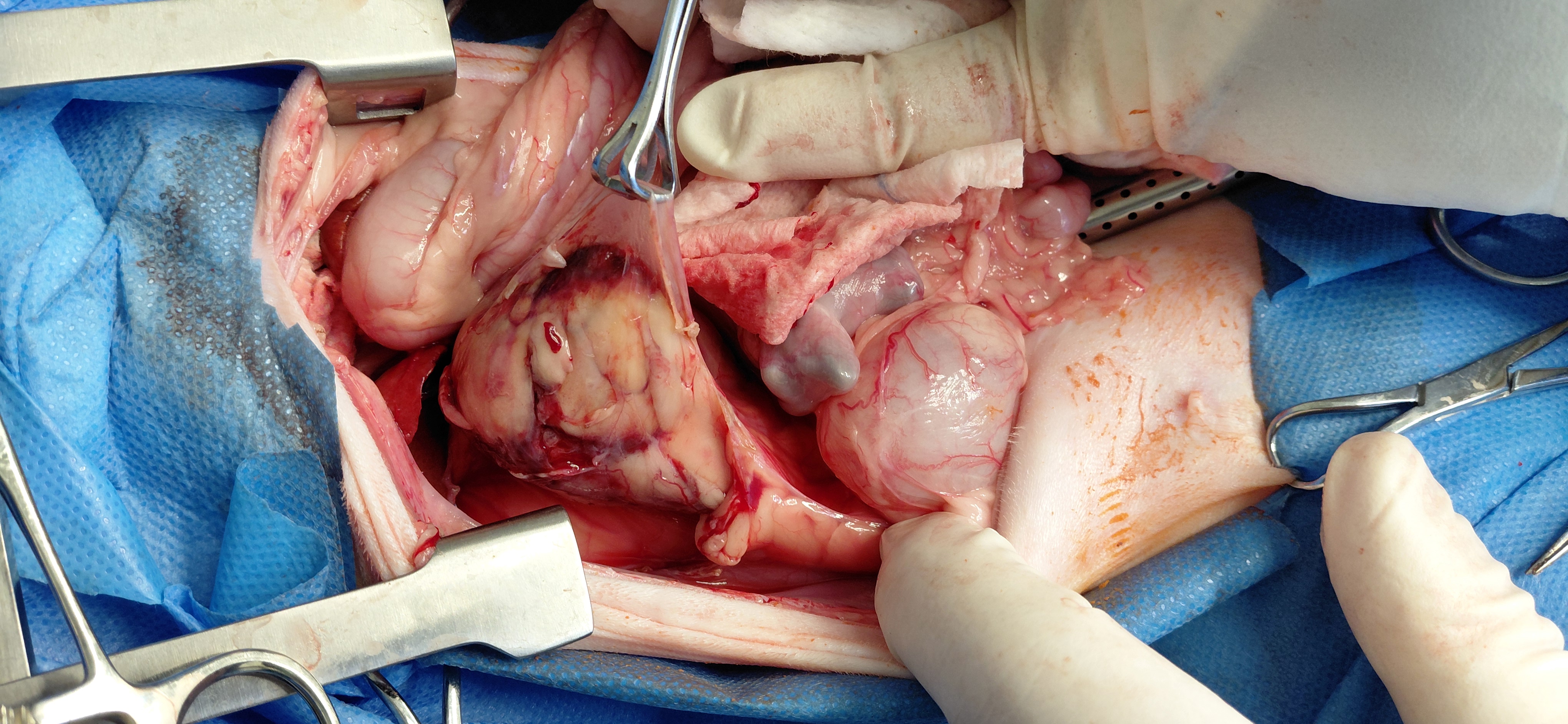
The abdomen was opened via a ventral midline incision. On opening, the team found a moderate amount of serosanguinous fluid (a mixture of blood and serum) already free within the abdomen, confirming that leakage had begun.
The extent of the disease was apparent immediately. The right ureter was markedly dilated, and the obstructing stone was palpable. The right kidney was severely enlarged and surrounded by inflamed, thickened perirenal fat that had adhered firmly to the surrounding abdominal wall - a consequence of the ongoing infection and inflammation. The blood supply to the kidney had increased dramatically in response, with extensive new vessel formation across the kidney surface.
The kidney was carefully dissected free from the retroperitoneum, with multiple small pericapsular vessels coagulated as the dissection proceeded. The renal artery was triple-ligated, including a central transfixation suture, and three branches of the renal vein were secured in the same way. The ureter was then dissected free from extensive adhesions along its full length, ligated just above the bladder, transected, and the ureteronephrectomy was complete.
After removal of the kidney and ureter, the abdomen was thoroughly lavaged and inspected carefully. A ligature was also placed on the phrenicoabdominal vein and on remnants of the ovarian vessels, which had been inadvertently damaged during the release of adhesions; something that can happen in cases where inflammation has altered the normal tissue planes significantly.
Abdominal closure was routine. Dolly recovered from anaesthesia and remained hospitalised for postoperative monitoring and intensive care.
Postoperative Care: The Days in Hospital
Dolly's pain management in hospital included methadone, transitioning to buprenorphine as she stabilised. She also received intravenous fluids, maropitant for nausea, marbofloxacin to continue tackling the infection, and felimazole to manage her hyperthyroidism. She began eating, and two days after surgery she was stable enough to go home.
Going Home: What Dolly's Owners Were Asked to Do
Caring for a cat after a nephrectomy (kidney removal) requires attention to several things at once: pain, infection, nausea, and protecting the remaining kidney from any additional stress.
Dolly's medications at home included buprenorphine transmucosal solution for pain, given under the tongue three times daily for three days; gabapentin oral solution three times daily for ten days; and marbofloxacin antibiotic tablets once daily. Her felimazole continued as before for the hyperthyroidism. The team also gave the option of bringing Dolly in for daily subcutaneous antibiotic injections if tablet administration proved too stressful (a practical acknowledgement that medicating cats is not always straightforward).
Exercise restriction and a calm home environment were advised throughout the recovery period.
What This Case Teaches Us
Dolly's case is a good example of how quickly obstructive kidney disease can escalate in cats and why prompt referral and decisive surgery can make the difference between survival and loss.
A few things stand out.
Subtle early signs matter
Weight loss and inappropriate urination are easy to attribute to age or behavioural change in a 14-year-old cat. Dolly's owners brought her in when they did, which gave the team a window to act before the kidney ruptured completely.
CT adds critical information
Ultrasound identified the mass and the hydronephrosis, but CT showed the precise location of the ureteral stone, the extent of perirenal involvement, and the early free fluid. All of which shaped the surgical plan directly.
Complex cases require a clear surgical priority
With adhesions, altered tissue planes, and a grossly enlarged kidney surrounded by inflamed fat, this was not a straightforward nephrectomy. The team's ability to manage intraoperative findings, including the incidental vessel damage during adhesion release, was as important as the primary procedure.
Age is not a contraindication
At 14 years old, Dolly was elderly and came in weak and hyperthyroid. But with appropriate pre-operative assessment, careful anaesthetic management, and comprehensive postoperative care, she came through surgery, began eating within 24 hours, and went home two days later.
"People sometimes assume that because a cat is elderly, surgery isn't the right path. But Dolly is a good example of why you have to look at the whole picture. Yes, she was 14. Yes, she was hyperthyroid and weak on arrival. But the alternative - leaving an infected, obstructed kidney in place - wasn't really an alternative at all. With careful anaesthetic planning and the right postoperative support, older cats can do remarkably well. Dolly proved that."
- Dr. Jakub Köcher-Vodnárek, Advanced Vetcare London
To refer a case or find out more about our surgical services, visit advancedvetcare.co.uk or call 020 3143 4444.