
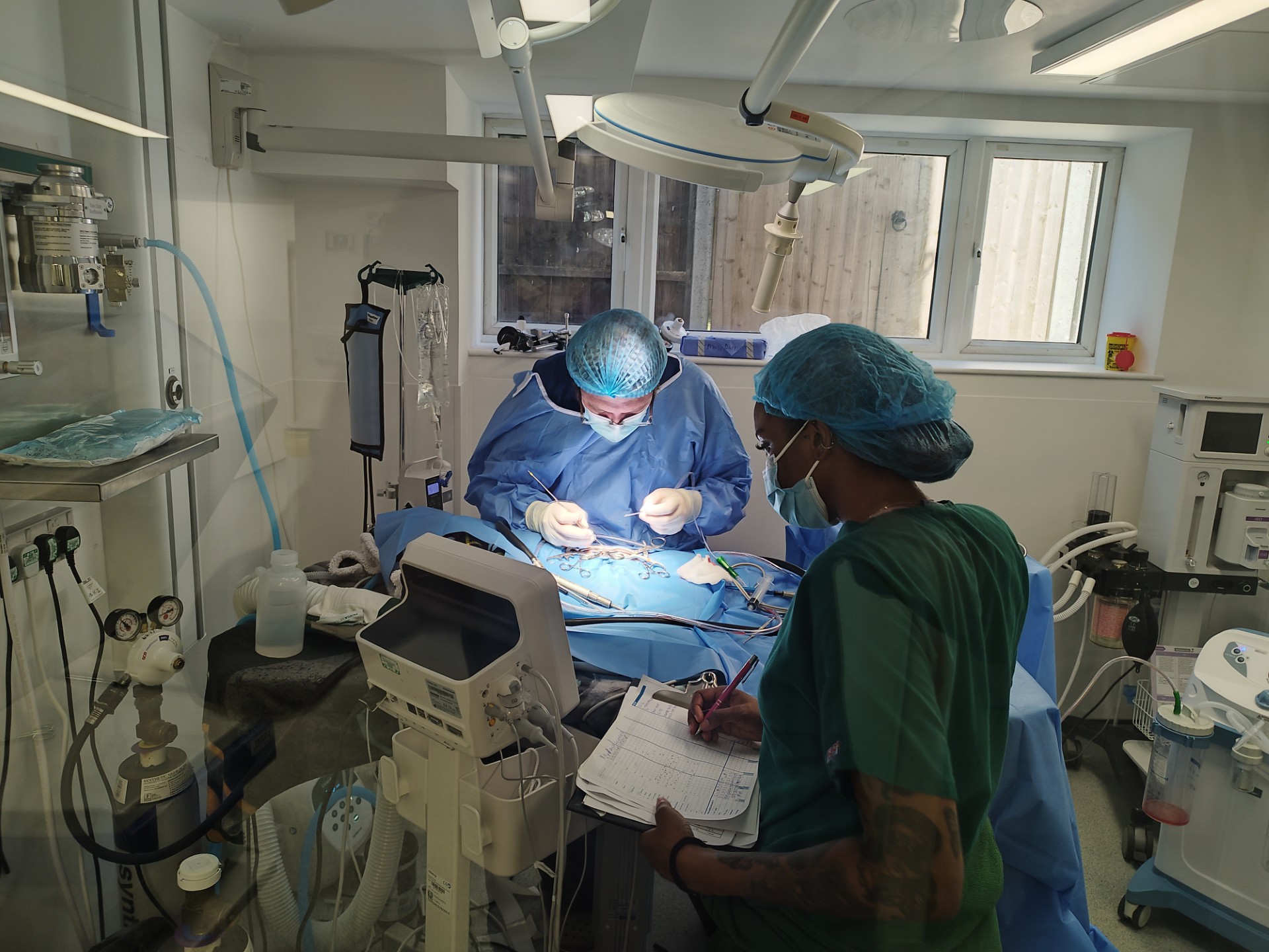
A technically demanding spinal surgery performed by Advanced Vetcare London in collaboration with London Veterinary Neurology.
When a “Wobbly Cat” Was Something More Serious
Sashi, a cat referred to Advanced Vetcare London, and managed in collaboration with London Veterinary Neurology, initially developed subtle mobility changes, including reluctance to jump and reduced agility around the home.
Over the following months, his condition worsened, and his owners noticed him deteriorating.
By the time of neurological assessment, Sashi was weak and struggling with balance and coordination. Although he remained able to walk, his neurological examination localised the problem to the thoracolumbar spinal cord (T3-L3 region). Notably, he showed minimal spinal pain, despite significant spinal cord compression.
This case highlights an important clinical lesson: a “wobbly cat” is not always inflammatory, infectious or neoplastic. Intervertebral disc disease, although uncommon in cats compared with dogs, is an important diagnosis in progressive feline mobility disorders.
Advanced Imaging Revealed a Severe Picture
Using advanced MRI imaging facilities provided by Advanced Vetcare London, South London’s only veterinary practice offering on-site pet MRI scanning facilities, clinicians identified a severe right-sided intervertebral disc lesion at T13-L1 causing marked spinal cord compression.
Initially, MRI alone could not definitively distinguish whether the lesion represented a disc extrusion, a disc protrusion, or a combination of both. However, surgery later confirmed the primary pathology was a large, firm, chronic intervertebral disc protrusion with only a small amount of extruded disc material.
A disc extrusion typically involves loose disc material within the vertebral canal, which can often be removed more readily during decompressive surgery. In contrast, a protrusion is usually firmer, more chronic and technically more difficult to access and remove safely.
In Sashi’s case, the protrusive nature of the lesion significantly increased the surgical complexity.
A More Active Approach Was Needed
Because Sashi was able to walk at the time of diagnosis, an initial period of conservative management was attempted. This included:
- Strict confinement
- Analgesia
- Careful neurological monitoring
However, due to the severity and chronicity of the spinal cord compression, clinicians considered long-term success with conservative management unlikely.
Over the following days, Sashi deteriorated further. His pelvic limb weakness worsened, particularly on the right side. He also began developing urinary and faecal retention. This represented a major clinical escalation and prompted urgent surgical intervention.
This case highlights an important point for veterinary surgeons: even cats with severe thoracolumbar spinal disease may benefit from surgery, even if their mobility is badly affected. However, the development of urinary or faecal problems should prompt urgent reassessment and referral discussion.
Dr. Jakub Köcher-Vodnárek, Veterinary Surgeon at AVL, said: “Sashi’s case was one of the most technically demanding feline spinal procedures I’ve assisted with. The close collaboration between Advanced Vetcare London and London Veterinary Neurology allowed Sashi to receive highly specialised surgical and nursing care throughout his treatment. Seeing him regain confidence, mobility and comfort after such severe spinal cord compression was incredibly rewarding for the whole team.”
A Rare and Technically Demanding Spinal Surgery
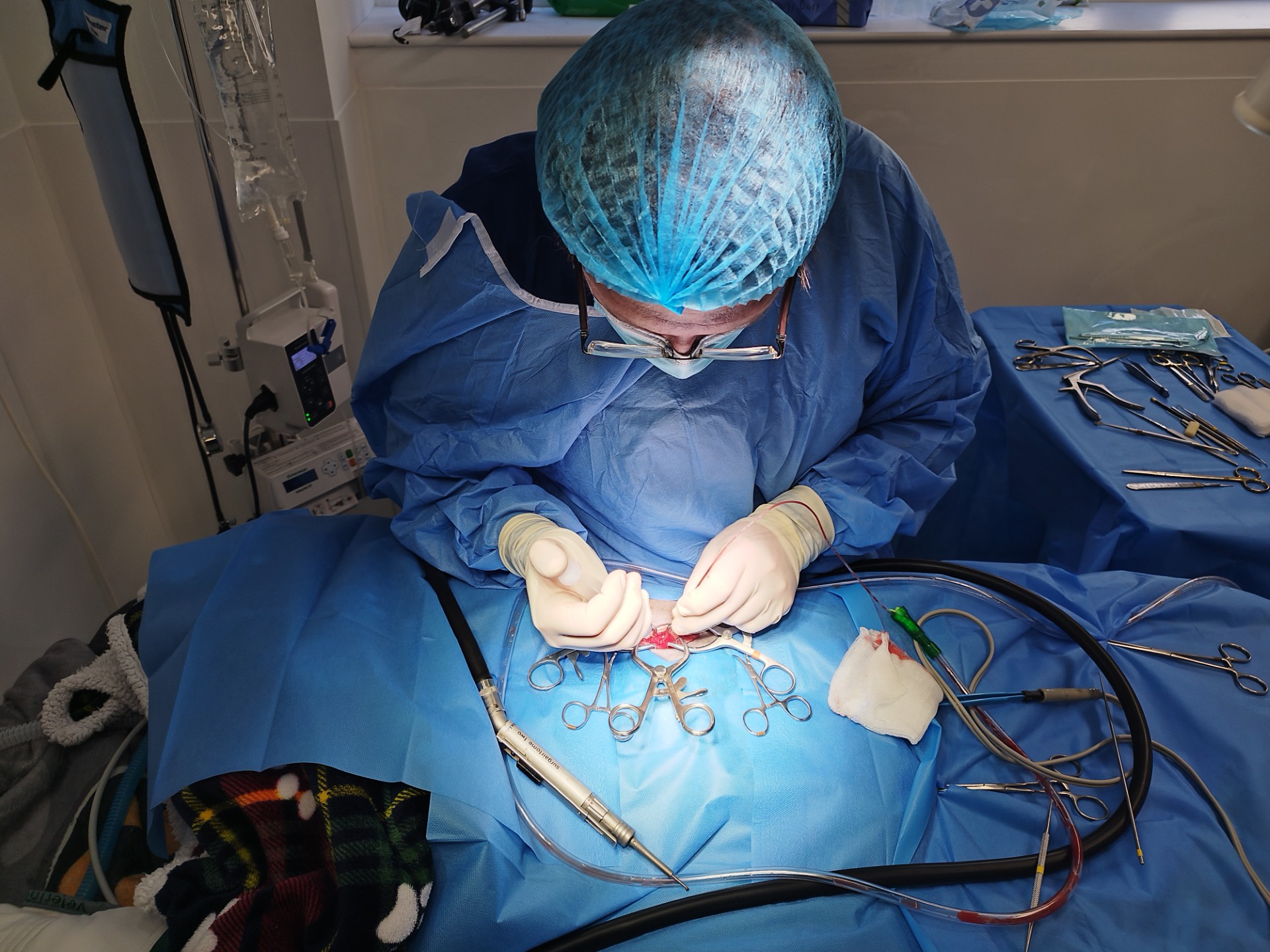
Sashi underwent a right-sided pediculectomy and corpectomy at T13-L1, performed by London Veterinary Neurology with perioperative support from Advanced Vetcare London.
A pediculectomy allows surgical access to the vertebral canal while preserving the articular facet. A corpectomy involves removing part of the vertebral body itself to access compressive material located beneath or beside the spinal cord.
This approach is uncommon in cats and is typically reserved for particularly difficult lesions, such as chronic, firm protrusions that cannot be adequately decompressed using more conventional techniques.
During surgery, the protruding disc was found to be causing severe spinal cord compression and displacement. The compressive material was carefully removed, allowing the spinal cord to return to a more normal anatomical position. No residual compression remained following decompression.

Challenges and Complications
The surgery was technically challenging.
The protruding disc had displaced both the epidural venous sinus and a nerve root directly into the surgical field. During decompression, the displaced nerve root was transected, resulting in a small focal cerebrospinal fluid (CSF) leak from the nerve-root sleeve.
Importantly:
- No central dural defect was identified
- No direct spinal cord injury was visible
- Adequate decompression could not have been safely achieved while preserving the displaced nerve root
Moderate venous haemorrhage was also encountered intraoperatively and controlled successfully.
This is a recognised risk in feline corpectomy procedures. As cats are small patients, the surgical corridor is extremely narrow, and even moderate blood loss can become significant.
What Happened During Post-Operative Care
Immediately following surgery, Sashi experienced transient neurological deterioration. However, his subsequent improvement and relatively short duration of worsening suggested this was more likely related to temporary post-decompression spinal cord dysfunction following chronic compression, rather than direct surgical spinal cord injury.
Post-operative management focused on:
- Strict confinement
- Intensive nursing support
- Analgesia
- Bladder monitoring
- Prevention of uncontrolled movement or jumping
Because Sashi had already developed urinary dysfunction prior to surgery, bladder function was monitored closely throughout recovery.
He remained under strict activity restriction for six weeks to allow healing and minimise the risk of postoperative reinjury.
Going Home with A Positive Recovery Ahead
Recovery following severe chronic spinal cord compression is often gradual and unpredictable. Some patients may initially appear unchanged, or even temporarily worse, during the first 24-48 hours after decompression before meaningful improvement becomes visible.
Fortunately, Sashi’s deterioration was short. He subsequently became brighter, more comfortable and increasingly functional.
Improvements were seen in:
- Standing ability
- Turning and balance
- Posture
- Managing toileting needs
- Overall demeanour
Although residual pelvic limb weakness and incoordination remain present, his current recovery path is positive.
What Makes This Case Study Stand Out?
- Feline intervertebral disc disease is uncommon but clinically important
- Severe spinal cord compression may occur even without marked spinal pain
- Disc protrusion and disc extrusion are not equivalent conditions surgically
- Chronic protrusions may require advanced decompressive procedures such as corpectomy
- Cats who are borderline non-ambulatory can still achieve meaningful recovery following surgery
- Urinary or faecal retention in thoracolumbar myelopathy is a major escalation sign
- Feline corpectomy remains a rare, technically demanding and high-risk procedure
A Collaborative Approach to Advanced Veterinary Care
Sashi’s case demonstrates the importance of early referral, advanced imaging, specialist surgical expertise and high-level nursing support in the management of severe neurological disease.
Through collaboration between Advanced Vetcare London and London Veterinary Neurology, Sashi was able to access advanced diagnostics, intensive perioperative care and a complex decompressive spinal procedure that offered the potential for meaningful neurological recovery.
Despite substantial deterioration prior to surgery, Sashi’s outcome remains encouraging, highlighting that even severe feline spinal cord compression may still be treatable when managed with timely referral and specialist intervention.
References
Amey JA, Liatis T, Cherubini GB, De Decker S, Foreman MH. Outcomes of surgically and conservatively managed thoracolumbar and lumbosacral intervertebral disc herniations in cats. Journal of Veterinary Internal Medicine. 2024;38(1):247-257.
Tyroller F, Wennemuth J, Forterre F, Flegel T, Markert C, Kiefer I, Wunderlin N. Retrospective study of partial lateral corpectomy to treat thoracic and lumbar intervertebral disc herniation in 12 cats. Journal of Feline Medicine and Surgery. 2024;26(12).