
Penny, a young dog, was referred to Advanced Vetcare London after a sudden injury left her unable to bear weight on her right foreleg. X-rays confirmed a fracture of both the radius and ulna, and specialist surgery was needed to give her the best chance of a full recovery.
How It Started
Penny's owners didn't see it happen. They were in another room when they heard her cry out, and when they went to find her, the reason was immediately obvious. She was holding her right foreleg completely off the ground, the lower portion of the limb hanging in a way that made it clear something was seriously wrong.
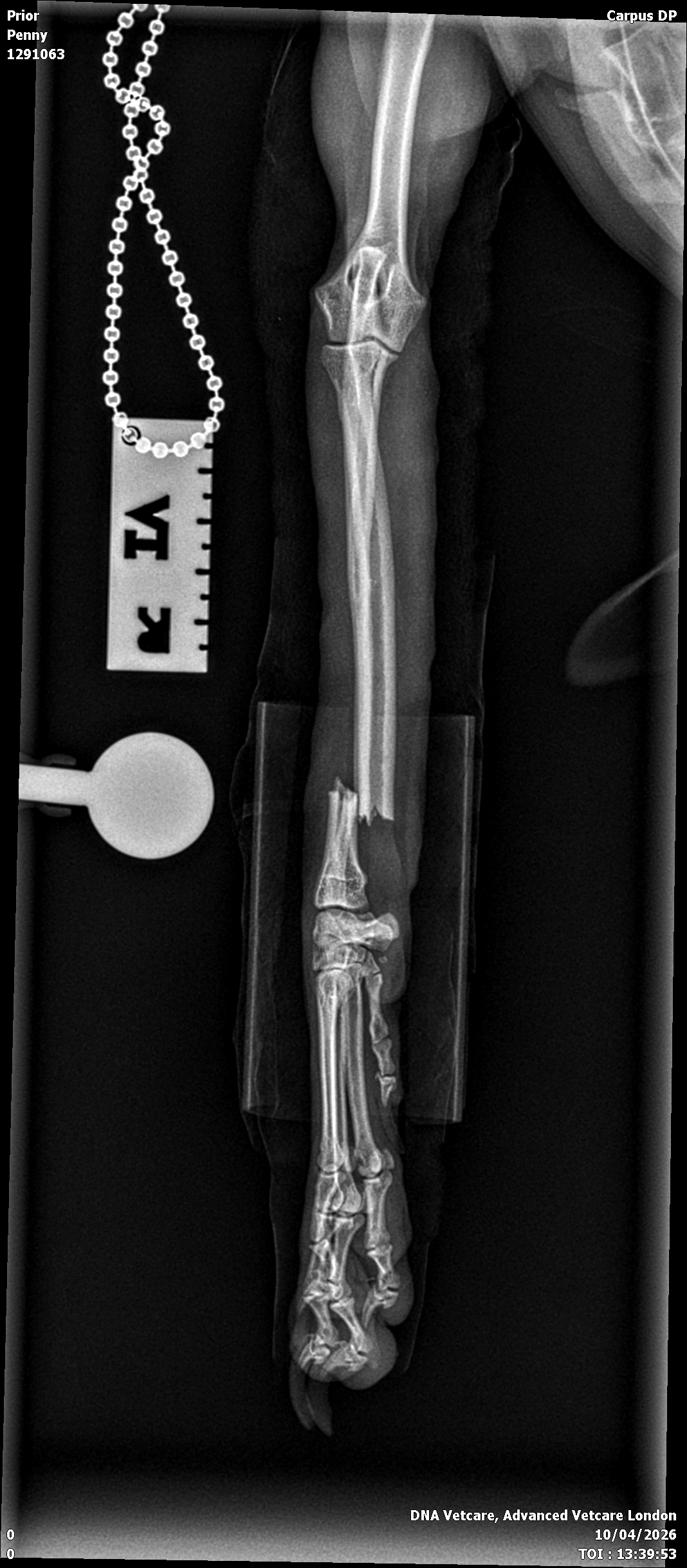
She was taken straight to Streatham Hills 24Hr Vetcare, where X-rays confirmed what the owners feared - a fracture of both the radius and ulna, the two bones that make up the lower forelimb. Penny was kept overnight on pain relief and cage rest before being referred to Advanced Vetcare London for specialist orthopaedic management.
The Fracture
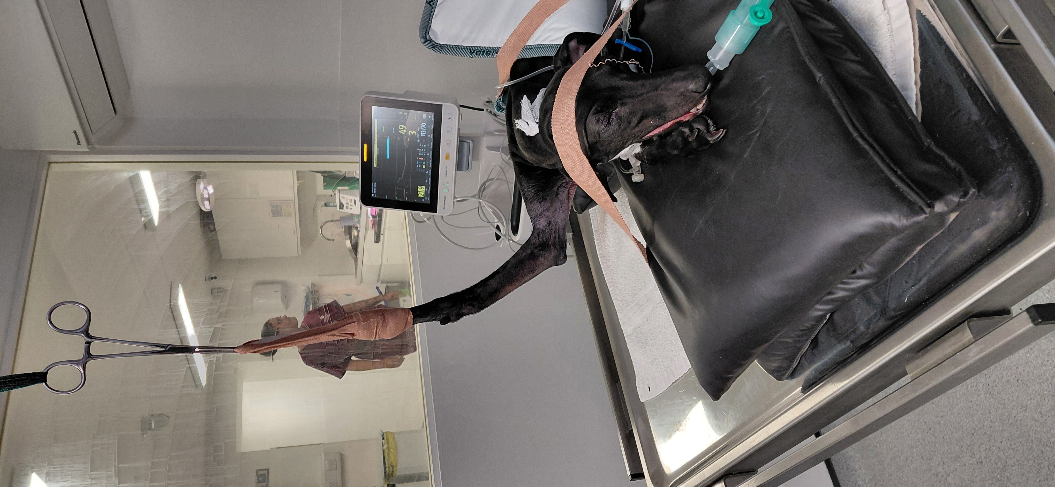
When Penny arrived the following morning, she was bright and alert - clearly still herself, despite the injury. Her vital signs were normal, and she had a bandage on the affected leg to keep things stable ahead of surgery.
Comparative orthogonal radiographs (X-rays taken from two angles at right angles to each other) confirmed a transverse fracture through the distal diaphysis - in plain terms, a clean crossways break near the lower end of both bones in the foreleg. This type of fracture, while serious, is well suited to surgical stabilisation using a bone plate, which is exactly the approach the team planned.
The Surgery
Penny was sedated and placed under general anaesthesia, with isoflurane used to maintain her throughout the procedure.
The surgical approach used was craniomedial - accessing the fracture site from the front and inner aspect of the foreleg, which gives the best exposure of the radius in dogs.
Once the fracture site was visible, the fragments were carefully reduced (brought back into their correct alignment) and held in position while fixation was applied.
A pre-contoured 10-hole 2.0 mm locking compression plate (LCP) was applied to the radius. These plates are designed to conform closely to the natural shape of the bone, and the combination of locking and compression screws used here provides stable, rigid fixation that protects the fracture while it heals.
Two cortical screws were placed in compression across the fracture site, drawing the two bone ends together, while three locking screws were placed in each fragment above and below the break. Locking screws anchor into the plate itself rather than relying purely on friction against the bone, which makes the overall construct considerably more robust.
The surgical site was thoroughly washed out before closure, and the wound was closed in layers (fascia, subcutaneous tissue, and skin) using a combination of absorbable and non-absorbable sutures depending on the layer and location.
Post-operative X-rays confirmed the fracture had been reduced well, with good alignment and implant positioning throughout.
"A distal radius and ulna fracture in a dog like Penny needs rigid, stable fixation to heal well. The locking compression plate gives us exactly that; it holds everything in the right position while the bone does its job of knitting back together. Penny's fracture reduced really nicely, and the post-operative films confirmed we were happy with the alignment throughout,” explains Mr. Jakub Köcher-Vodnárek, Diplomate ECVS, Advanced Vetcare London.

After Surgery
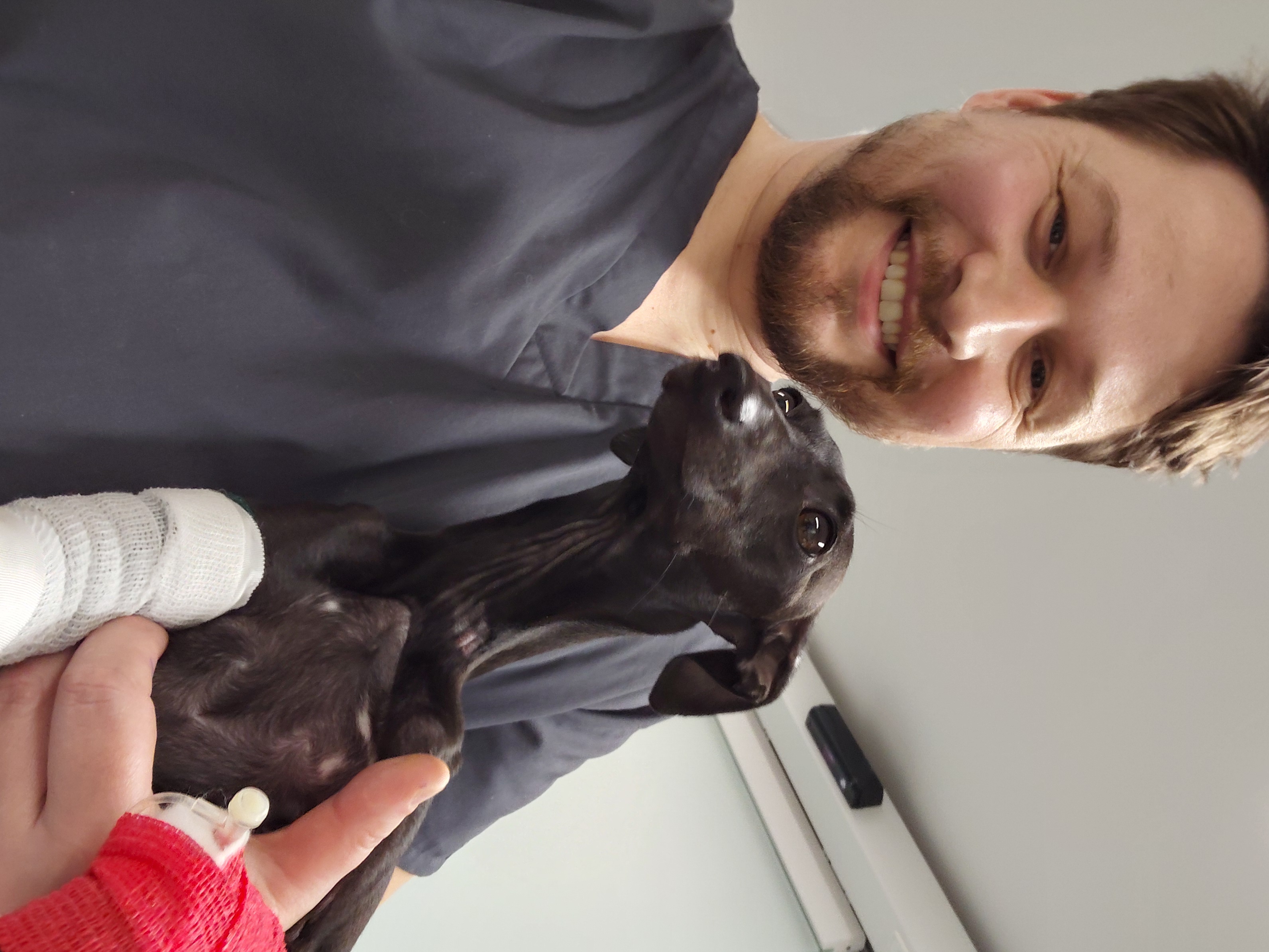
Penny came through the procedure without any complications and recovered from the anaesthetic smoothly. A light bandage was applied to the leg and toes left exposed at the bottom so circulation could be checked, and stopping just below the elbow at the top.
Penny stayed in hospital for one night at Streatham Hills Veterinary Surgery and, once she had recovered well, was returned to her owners the following day with a detailed plan for the weeks ahead.
The Road Home
The next 8 weeks are the most important part of Penny's recovery. Strict cage or pen rest is essential during that time. The plate does the job of stabilising the fracture, but bone healing takes time, and too much activity too soon risks disrupting that process.
Her bandage will be changed regularly, and sutures will come out at the two-week mark. The bandage itself is likely to stay on for 6 to 8 weeks in total, depending on how things progress.
Controlled lead exercise can begin in the first fortnight - short walks of 5 to 10 minutes, three times a day, with gradual increases of 5 minutes per week after that. A harness rather than a collar is recommended to take the load off the injured limb during walks, and weight-balancing exercises at home can start from week one to help Penny begin rebuilding strength and confidence in the leg.
Follow-up X-rays at 6 to 8 weeks will allow the team to assess fracture healing and check for stress shielding. This is a process where the bone can become less dense under the plate if it isn't bearing load properly. Penny will need a short sedation for those images, so owners will need to fast her beforehand.
What This Case Teaches Us
Penny's case is a good example of why prompt, specialist management of limb fractures makes such a difference in dogs. The fracture involved both bones of the foreleg, which means stability couldn't come from one bone compensating for the other - surgical fixation was the right call, and the LCP plating system used here is well established as the gold standard for this type of injury.
It also highlights the value of the referral pathway. Streatham Hills 24Hr Vetcare stabilised and managed Penny's pain overnight, which meant she arrived at Advanced Vetcare London in good condition and ready for surgery. That kind of joined-up care between first-opinion and specialist teams makes a real difference to how smoothly a case like this unfolds and ultimately, to the outcome for the patient.